


Point-of-care ultrasound (POCUS) is an ultrasound examination performed at the point of care, interpreted in real time, and integrated into the clinical context by the treating team. It is usually designed to answer a focused question—not to survey every possible abnormality. Used well, POCUS connects a clinical question to image acquisition, interpretation, and the next appropriate step. Used without adequate training or outside its limits, it can create false reassurance or lead to incorrect conclusions.
Quick answer: POCUS is best understood as a focused clinical tool and a repeatable workflow. It may complement the physical examination, guide procedures, monitor change, or help determine whether additional testing is needed. Whether it can replace another imaging study depends on the clinical question, the quality and scope of the examination, the operator’s training, and local standards.
What is POCUS?
The clearest current definition comes from a multidisciplinary expert consensus: POCUS is performed at the point of care, interpreted and integrated into context by the treating team, and typically has a targeted scope focused on a specific clinical question. The same consensus notes that POCUS can help identify, guide, or evaluate procedures as well as support diagnostic and monitoring tasks. See the full 2024 interdisciplinary POCUS best-practices consensus.
The American Institute of Ultrasound in Medicine (AIUM) similarly describes a POCUS examination as a unique diagnostic test that is complementary to the physical examination. Its practice parameter for point-of-care ultrasound examinations emphasizes that not every abnormality can be detected and that an additional or comprehensive ultrasound examination may be necessary.
POCUS therefore is not simply a small ultrasound machine, a handheld device, or a collection of memorized images. It is a clinical process:
- Start with an appropriate, focused question.
- Acquire the required views with adequate image quality.
- Interpret the images within the limits of the examination.
- Integrate the result with the history, physical examination, laboratory data, and other imaging.
- Document the study and escalate when the question remains unanswered.
POCUS versus comprehensive ultrasound
POCUS and comprehensive ultrasound can examine the same anatomy, but they are not automatically interchangeable. A focused cardiac examination, for example, is not synonymous with a comprehensive echocardiogram. The correct comparison is based on scope, workflow, training, and the question being asked—not simply the device used.
| Feature | POCUS | Comprehensive or consultative ultrasound |
|---|---|---|
| Primary purpose | Usually answers one or a limited set of focused clinical questions | Performs a broader, standardized evaluation of an organ or region |
| Who integrates the result | The treating clinical team, often during the encounter | An imaging specialist or dedicated laboratory communicates a formal interpretation |
| Timing | Often immediate and repeatable at the bedside | Usually scheduled or requested through an imaging workflow |
| Scope | Targeted and application-specific | More detailed and comprehensive for the requested examination |
| Documentation | Representative images or clips plus a clinical note, according to local policy | Standard image set and formal report according to laboratory standards |
| Can it replace the other? | Sometimes, but not by default. The answer depends on the clinical context, image quality, operator competence, scope, findings, and local standards. | |
The expert consensus states that POCUS tends to provide more immediate data and is easier to repeat, but in most cases is less detailed and comprehensive. A successful POCUS examination may or may not remove the need for additional imaging. The New England Journal of Medicine review of POCUS likewise describes POCUS and consultative ultrasonography as complementary.
The I-AIM workflow: a reliable way to think through every scan
The I-AIM model organizes focused sonography into four connected domains: indication, acquisition, interpretation, and medical decision-making. It was proposed as both a mnemonic and a checklist in the original I-AIM publication. It is useful because it prevents the image from becoming disconnected from the clinical question.
1. Indication: What question are you trying to answer?
Define the question before selecting the probe. A focused question should be relevant to the clinical presentation and within the operator’s scope. Consider the pretest context, the strengths and limitations of the chosen examination, and what you will do if the images are inadequate, negative, positive, or indeterminate.
“Perform a POCUS exam” is not a complete indication. “Use a locally approved focused protocol to assess a defined question in this clinical context” is a more disciplined starting point.
2. Acquisition: Can you obtain the required views?
Acquisition includes patient positioning, probe and preset selection, orientation, standard views, systematic sweeping, and image optimization. An image that contains the target is not necessarily an adequate examination. Each application has required anatomy, views, and quality criteria that should be learned from specialty-specific guidance and supervised practice.
3. Interpretation: Is the examination adequate, and what do the images show?
Interpretation starts by judging technical adequacy. Identify normal landmarks, artifacts, expected variants, and the limits of the view before assigning a clinical meaning. “Indeterminate” is an appropriate interpretation when the necessary anatomy is not visualized or the finding remains uncertain.
4. Medical decision-making: How does the result fit the whole patient?
POCUS findings should be integrated with the history, examination, vital signs, laboratory results, and other testing. A focused study is one data source, not an isolated verdict. If the result conflicts with the clinical picture, exceeds the examination’s validated scope, or does not answer the original question, further testing or expert review may be necessary.
For a short worked example, open the ED Room 6 preview. Choose a recorded view, save it, and return to that scan while answering one question. Notice what the image supports and what still needs clarification.
Common POCUS application families
POCUS is used across emergency medicine, critical care, hospital medicine, anesthesia, family medicine, pediatrics, and other settings. The exact scope differs by specialty and institution. The 2023 American College of Emergency Physicians ultrasound guidelines describe a broad emergency-ultrasound scope, while other societies publish recommendations tailored to their own clinicians and patient populations.
| Application family | Examples of focused tasks | Important boundary |
|---|---|---|
| Cardiac and hemodynamic | Focused assessment of selected cardiac structures or function in a defined context | Not automatically equivalent to a comprehensive echocardiogram; nomenclature, views, and competency expectations vary |
| Lung and pleural | Assessment for selected pleural or pulmonary patterns in an appropriate clinical setting | Artifacts must be interpreted in context, and technically limited examinations may require other imaging |
| eFAST and trauma | Protocol-based search for specific findings in trauma | A negative or incomplete focused examination does not exclude every injury |
| Vascular | Focused compression assessment or procedural guidance within an approved protocol | Protocol extent and follow-up pathways matter; comprehensive vascular imaging may still be needed |
| Abdominal and urinary | Focused aortic, biliary, renal, bladder, or free-fluid questions | Bowel gas, body habitus, pain, dressings, and incomplete visualization can limit the study |
| Soft tissue and musculoskeletal | Focused evaluation of superficial structures or dynamic anatomy | Angle-dependent artifacts and incomplete field of view can mislead an inexperienced operator |
| Procedural guidance | Identify anatomy, guide a device, or evaluate a procedure within training and policy | Requires procedure-specific training, sterile technique when indicated, and continuous awareness of device position |
This table is an educational map, not a protocol. Use the current guidance of the relevant specialty society and your institution before performing or interpreting any application independently.
Begin with a focused clinical question, select the appropriate examination, obtain adequate views, and interpret the findings in context. The application families below are not exhaustive; training, scope, and local protocols determine appropriate use.
Probes, presets, and imaging modes
Probe selection balances depth, image detail, footprint, and the available acoustic window. The common probe categories below are a starting point; exact bandwidth and capabilities differ by system.
| Probe | Typical characteristics | Common educational examples |
|---|---|---|
| Linear array | Higher frequency, rectangular image, strong near-field detail | Vascular, superficial soft tissue, musculoskeletal structures, and selected procedures |
| Curvilinear array | Lower frequency, broad curved footprint, deeper abdominal field | Abdominal, pelvic, renal, biliary, and trauma views |
| Phased array | Lower frequency, small footprint, sector-shaped image | Cardiac and intercostal windows |
Higher-frequency sound generally provides more detail with less penetration, while lower-frequency sound penetrates more deeply with less fine detail. That trade-off, along with pulse–echo imaging, gain, depth, focus, Doppler, and artifacts, is explained in 10 ultrasound physics principles for better POCUS images.
Common imaging modes
- B-mode: the standard two-dimensional grayscale image.
- M-mode: displays motion along one ultrasound line over time.
- Color Doppler: maps Doppler information over a B-mode image according to the selected scale and color map.
- Spectral Doppler: displays Doppler frequency information over time from a defined sampling method.
Selecting a mode is not a substitute for knowing its assumptions, artifacts, and limitations. Learn application-specific Doppler techniques under qualified instruction before using measurements for clinical decisions.
Orientation and probe movement
Every ultrasound transducer has an orientation marker, and the screen has a corresponding marker. Conventions can differ among specialties, particularly in cardiac imaging. The AIUM practice parameter explicitly notes that different subspecialties place the marker on different sides of the screen. The practical rule is to confirm the convention used in your curriculum and clinical environment, then apply it consistently.
Most image acquisition can be described with a small movement vocabulary:
- Slide: move the entire probe across the skin while maintaining its orientation.
- Rotate: turn the probe around its central axis.
- Rock: move the beam along the probe’s long axis while maintaining the contact point.
- Tilt or fan: angle the beam across the short axis while maintaining the contact point.
- Sweep: scan through the target to evaluate its full extent rather than stopping at one frame.
- Compression: apply controlled pressure when the application calls for it and the patient can tolerate it.
Small, deliberate movements are easier to interpret than several simultaneous corrections. If the view deteriorates, return to the last recognizable landmark and change one variable at a time.
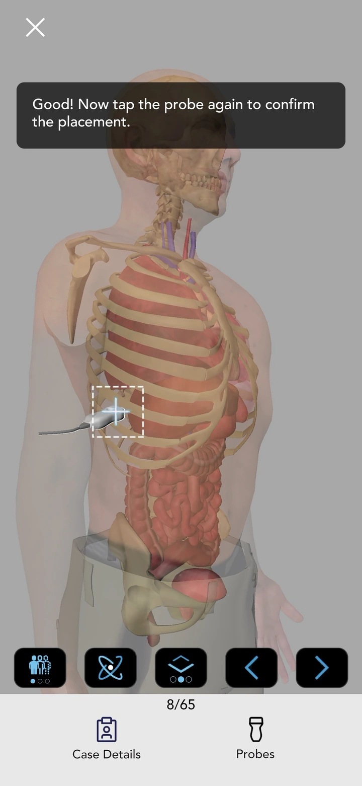
Image optimization: make the examination interpretable
Start with the correct probe and preset, then optimize the image in a repeatable sequence. The exact order may vary by application, but beginners benefit from a consistent checklist:
- Depth: include the full target without leaving excessive unused far field.
- Overall gain: adjust received-signal amplification so fluid, soft tissue, and strong interfaces are distinguishable.
- Time gain compensation: correct depth-dependent brightness only when needed.
- Focus: place the focal zone at or just beyond the target according to the system and application.
- Frequency: use the highest frequency that still reaches the target adequately.
- Patient and probe position: improve the acoustic window rather than relying only on controls.
Gain is not acoustic output. Increasing gain brightens amplified returning signals; increasing output changes transmitted acoustic energy. This distinction matters for both image quality and prudent exposure. The ACEP Sonoguide physics and machine-controls chapter provides an accessible introduction.
How to learn POCUS responsibly
There is no single universal training path for every profession, application, and clinical environment. The 2024 interdisciplinary consensus recommends a flexible framework with three major elements:
- Didactic learning: physics, probe selection, image optimization, artifacts, standard views, normal anatomy, and relevant pathology.
- Directly supervised hands-on practice: scanning models, volunteers, simulators, or consenting patients with immediate expert feedback.
- Monitored use: clinical scanning with ongoing review of archived images and interpretations as the learner transitions toward independent practice under local standards.
A mobile or web-based tool can support the first element and portions of interpretation and decision rehearsal. It cannot reproduce tissue pressure, ergonomics, live anatomy, patient variability, or supervised acquisition. The evidence review summarized in how a POCUS simulator app fits into ultrasound education explains what online learning can and cannot reasonably claim.
Competence is more than a scan count
Minimum examination numbers may be used within specialty pathways, but experience develops at different rates and different applications have different learning curves. A 2024 systematic review of POCUS psychomotor learning curves found substantial heterogeneity and a lack of clear, validated definitions for learning endpoints. Scan counts should therefore be combined with qualitative assessment of image acquisition, interpretation, and integration—not treated as proof of competence by themselves.
For a practical starting routine, continue with 10 evidence-informed POCUS habits for beginners.
Limitations: know when the answer is “not adequately assessed”
POCUS is operator-dependent. Its performance can be limited by body habitus, bowel gas, subcutaneous emphysema, pain, patient position, dressings, tubes, time pressure, and the operator’s experience. Disease can also lie outside the field, protocol, timing, or validated purpose of the focused examination.
The AIUM parameter recommends a comprehensive study when a limited examination does not answer the clinical question or when an incidental finding warrants further investigation. A disciplined operator should be prepared to record one of three broad outcomes:
- Adequate and interpretable: the required views were obtained and can be interpreted within the protocol’s scope.
- Technically limited: some required anatomy or views were not adequately obtained.
- Indeterminate or discordant: images may be adequate, but the result does not confidently answer the question or does not fit the clinical context.
Negative does not mean comprehensive, and technically limited does not mean normal. If uncertainty could affect care, seek expert input or the appropriate additional test.
Documentation, archiving, and quality assurance
POCUS is a clinical examination, so image storage and documentation should follow clinical standards. Exact requirements vary, but a robust system usually includes:
- The indication or focused question.
- Representative labeled still images or clips.
- The views obtained and any missing or limited views.
- The interpretation, including uncertainty and limitations.
- How the result was integrated into the plan.
- A pathway for expert review, feedback, and correction.
- Secure storage consistent with privacy and institutional policy.
The multidisciplinary consensus recommends supervisory or quality-assurance verification during training and recognizes ongoing expert review as a useful clinical-quality and educational process. Your institution’s policy determines which examinations require archiving, how they are reported, and who may review them.
Ultrasound safety and infection control
Diagnostic ultrasound does not use ionizing radiation, but that does not mean the correct safety message is “no risk.” Ultrasound transmits acoustic energy, and prudent use requires attention to output and exposure. AIUM recommends the as low as reasonably achievable (ALARA) principle: use the lowest acoustic output that provides the needed diagnostic information, monitor the displayed thermal and mechanical indices, avoid unnecessary stationary dwell time, and keep the examination only as long as needed. Review the current AIUM ALARA statement.
Infection control is equally important. The AIUM 2025 transducer and coupling-gel guidance states that external transducers require cleaning and low-level disinfection between patients, while internal probes require high-level disinfection and a single-use cover. Interventional and nonintact-skin situations have additional requirements. Always follow the transducer manufacturer’s instructions, applicable regulations, and local infection-control policy.
A practical path for getting started
- Choose one relevant application. Start with an examination that fits your clinical role and has a defined local curriculum.
- Learn the question and limits. Know what the examination is designed to answer and what it cannot exclude.
- Study normal anatomy and standard views. Build landmarks before focusing on pathology. Want one structured reference beside your training? Match a current POCUS book to your level and specialty.
- Rehearse the sequence. Use diagrams, saved clips, cases, or simulation to practice probe choice, protocol order, interpretation, and decision logic.
- Scan under direct supervision. Obtain immediate feedback on ergonomics, probe movement, image quality, and interpretation.
- Save and review your studies. Track technical errors and feedback, not just totals.
- Complete local assessment and credentialing. Meet the standards for your role, specialty, and institution before independent use.
- Continue quality review. Maintain skills through use, feedback, case review, and continuing education.
If you need supervised scanning opportunities, browse the Ultrasound Fanatic directory of POCUS workshops and ultrasound events. Verify course scope, faculty, accreditation, prerequisites, and current registration details on each organizer’s official page.
Frequently asked questions about POCUS
What does POCUS stand for?
POCUS stands for point-of-care ultrasound or point-of-care ultrasonography. It generally refers to an ultrasound examination acquired, interpreted, and integrated into the clinical context by the treating team at the point of care.
Is POCUS the same as a formal ultrasound or echocardiogram?
Not necessarily. POCUS usually has a more focused scope and an immediate bedside workflow. A comprehensive ultrasound or echocardiogram follows its own acquisition, measurement, reporting, and specialist-interpretation standards. A POCUS study may sometimes answer the question sufficiently, but in other situations additional imaging remains necessary.
Who can perform POCUS?
Different regulated health professionals perform POCUS within their education, licensure, specialty scope, institutional privileges, and local law. There is no single universal answer. Training and authority for one application do not automatically confer competence or permission for every other application.
How long does it take to learn POCUS?
There is no universal timeline. Learning curves differ among applications and learners. Competence requires more than completing a course or reaching a scan count; it includes appropriate indication, adequate acquisition, accurate interpretation, clinical integration, and demonstrated performance under the relevant local standard.
Can a normal POCUS examination rule out every relevant condition?
No. A focused examination is limited to its protocol, views, image quality, timing, operator performance, and validated scope. A negative or technically limited POCUS examination should not be generalized beyond what was adequately assessed.
Can I learn POCUS without owning an ultrasound machine?
You can learn physics, anatomy, protocol structure, probe-selection concepts, and image interpretation without owning a machine. Simulation and online cases can support that preparation. Real image-acquisition skill still requires hands-on practice with an ultrasound system, appropriate models or patients, and qualified supervision.
Does POCUS use radiation?
POCUS does not use ionizing radiation. It uses acoustic energy. Operators should still practice ALARA, monitor output indices as appropriate, and keep exposure only as long and as high as needed to obtain useful information.
Selected authoritative references
- Oto B, et al. Best Practices for Point of Care Ultrasound: An Interdisciplinary Expert Consensus. 2024.
- AIUM Practice Parameter for the Performance of Point-of-Care Ultrasound Examinations.
- American College of Emergency Physicians. Ultrasound Guidelines: Emergency, Point-of-Care, and Clinical Ultrasound Guidelines in Medicine. 2023.
- Díaz-Gómez JL, Mayo PH, Koenig SJ. Point-of-Care Ultrasonography. New England Journal of Medicine. 2021.
- Bahner DP, Hughes D, Royall NA. I-AIM: A Novel Model for Teaching and Performing Focused Sonography. 2012.
- Breunig M, Chelf C, Kashiwagi D. Point-of-Care Ultrasound Psychomotor Learning Curves: A Systematic Review. 2024.
- AIUM. As Low As Reasonably Achievable (ALARA) Principle.
- AIUM. Guidelines for Cleaning Ultrasound Transducers and Safe Handling of Coupling Gel. 2025 revision.


