


Short answer: On lung ultrasound, A-lines are repeating horizontal reverberation artifacts below the pleural line. B-lines are bright vertical artifacts that arise from the pleural line, classically extend into the far field and interrupt or erase A-lines along their path. Their appearance depends on the imaging settings. A-lines usually reflect aerated lung at the scanned location; B-lines reflect a change in the air–fluid or air–tissue mixture near the pleural surface. Neither pattern is a diagnosis by itself.
Interpretation depends on image quality, lung sliding, the pleural line, the number and distribution of artifacts, other lung-ultrasound findings, and the complete clinical context. “A-lines equal normal” and “B-lines equal pulmonary edema” are useful memory shortcuts only until they become unsafe oversimplifications.
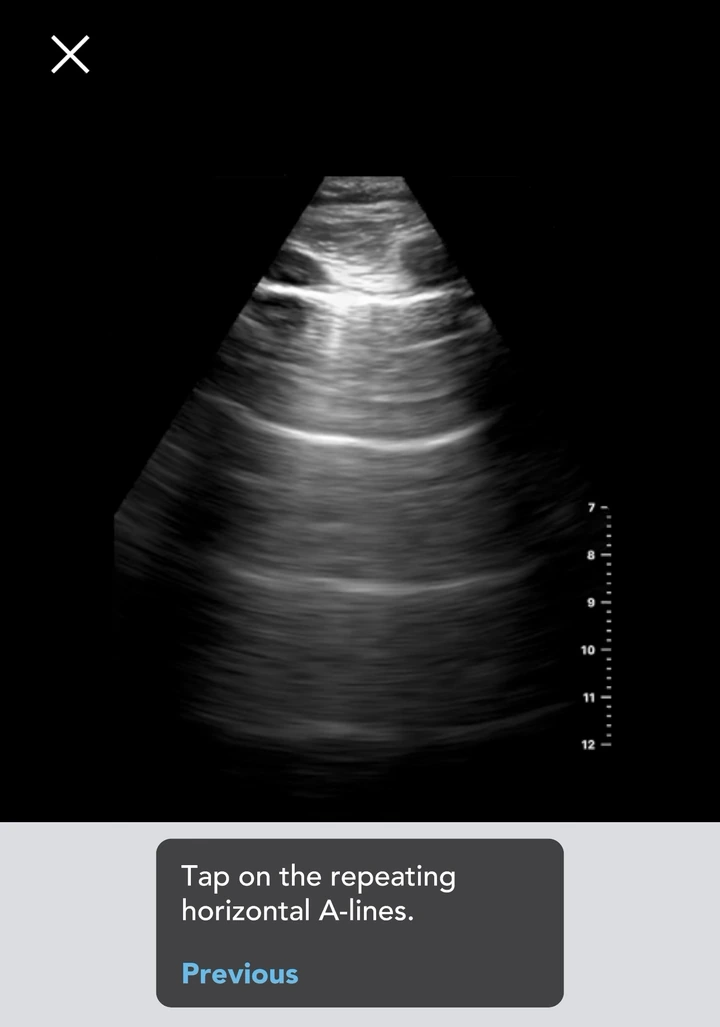
A-line reference example
B-line reference example

A-lines vs B-lines: the quick comparison
| Feature | A-lines | B-lines |
|---|---|---|
| Direction | Horizontal | Vertical |
| Origin | Repeating reverberation of the pleural line | Arise from the pleural line |
| Depth behavior | Repeat at regular intervals related to the probe-to-pleura distance and usually become less bright with depth | Classically persist into the far field; displayed length depends on settings |
| Relationship to A-lines | They are the repeating horizontal pattern | Erase or interrupt A-lines along their path |
| Respiratory movement | The lines themselves are generally static; assess the pleural line separately for sliding | Move with the pleural line when lung sliding is present |
| What the pattern supports | Aerated lung beneath the scanned location | Reduced aeration or increased density near the pleural surface |
| Main pitfall | A-lines can occur in both normal and abnormal clinical states | B-lines have multiple causes and are sensitive to technique and settings |
The one-sentence distinction: A-lines are horizontal repeats of the pleural line; B-lines are vertical artifacts from the pleural line that reach the far field and erase A-lines along their course.
See A-lines and B-lines in motion
Watch how the app asks a learner to identify A-lines and B-lines in moving reference images. The recorded app images and explanations in this guide help you study the patterns alongside these examples.
These excerpts use separate reference examples, not a same-patient comparison. Use the image-quality, distribution and clinical-context checks in this guide when interpreting the patterns.
Read the recording description
The first excerpt shows the A-line identification example in the app’s Normal Lung module. The second shows a B-line target in the Interstitial module, a learner tap and the accepted feedback. The clinical images and app responses are unchanged.
Original POCUS Simulator app recording. Educational recognition practice, not a complete examination.
Check your reasoning in 60 seconds
After watching the clip, answer these three learning prompts before opening each explanation. Name the finding, then state its limit.
1. Is a bright vertical streak alone enough to identify a B-line?
No. Check its pleural origin, relationship to A-lines, and motion in a cine clip. Length alone is not a universal pass/fail test: depth and other settings alter artifact appearance. Review the settings caveat below.
2. A still frame shows repeating A-lines. Can you conclude that lung sliding is present?
No. A-lines describe an artifact pattern; lung sliding is motion over time. Review a cine clip or the live image to assess sliding. Keep these observations separate: an A-line pattern by itself does not establish that the lung is normal.
3. You recognize B-lines in one view. Does that establish pulmonary edema?
No. B-lines are not specific to a single disease. First describe image quality and the pattern at that location. Its meaning depends on distribution across the examination, the pleural line, associated findings, and the clinical context.
If you missed one, return to the comparison table and explain the distinction in your own words. These prompts rehearse concepts; they are not a clinical competency assessment. Criteria: ACEP Sonoguide and Lung Ultrasound: The Essentials.
Start with the pleural line, not the artifact
Lung ultrasound is different from many other ultrasound applications because much of the information comes from artifacts created at the chest wall–pleural–lung interface. Air scatters and reflects ultrasound strongly, so a normally aerated lung is not displayed as a detailed picture of the lung parenchyma. Instead, the operator evaluates the pleural line, its motion, and the artifact pattern beneath it.
First locate an intercostal space. The ribs appear as bright curved interfaces with posterior acoustic shadows. Between them, the closely apposed parietal and visceral pleura usually appear as one bright horizontal line. This rib–pleural-line arrangement is often called the bat sign.
The ACEP Sonoguide lung chapter emphasizes that accurate lung-ultrasound interpretation depends on recognizing these artifacts and their relationship to the pleural interface. If the pleural line is not confidently located, do not label a bright streak as an A-line or B-line yet.
A quick image-quality check
- Are one or both rib shadows visible?
- Is the pleural line identified between the ribs rather than confused with a superficial fascial line?
- Is the depth sufficient to show the expected far-field behavior of the artifact?
- Is the gain balanced rather than washing out subtle lines or manufacturing excess brightness?
- Is the beam reasonably perpendicular to the pleural surface?
- Is this a moving clip when pleural motion is part of the question?
A recent Society of Hospital Medicine position statement on POCUS image quality includes the pleural line, rib shadows, balanced gain, sufficient depth, and perpendicular beam alignment among the quality elements used for common lung images. An artifact should not be interpreted separately from the quality of the view that produced it.
What are A-lines on lung ultrasound?
A-lines are horizontal reverberation artifacts that appear beneath the pleural line. They occur because sound reflects repeatedly between the transducer and the strong pleural–air interface. The ultrasound system places each delayed returning echo deeper in the image, creating several copies of the pleural line.
This produces three useful recognition features:
- A-lines are parallel to the pleural line.
- The interval between successive A-lines relates to the distance from the transducer to the pleural line.
- The repeated lines often become less bright with depth as energy is lost.
The RSNA review Lung Ultrasound: The Essentials explains that A-lines allow the examiner to infer aerated lung beneath the pleural interface. That is a description of the artifact at one scanned location—not a conclusion that the entire lung or patient is normal.
Do A-lines mean normal lung?
Not automatically. A-lines commonly predominate over normally aerated lung, especially when accompanied by a regular pleural line and lung sliding. They may also appear in clinical states where the lung remains aerated, including obstructive disease, and they can be present when pleural air prevents the visceral and parietal pleura from remaining apposed.
That is why an A-line pattern must be described with other observations:
- Was lung sliding present, absent, or not confidently assessed?
- Was the pleural line regular or abnormal?
- Were the findings focal or widespread?
- Were consolidations, pleural fluid, or other signs present?
- Was the examination technically adequate and complete for the focused question?
Safe wording: “An A-line-predominant pattern is present at this location” is more accurate than “the lung is normal.”
What are B-lines on lung ultrasound?
B-lines are bright vertical artifacts associated with a change in the mixture of air and fluid or tissue near the pleural surface. Their exact physical origin is complex and remains an active area of research, so it is safer to recognize the established image features than to rely on one simplified physics explanation.
These are the classic recognition features, interpreted together rather than as a universal device-independent checklist:
- It arises from the pleural line.
- It is bright and predominantly vertical.
- It classically persists into the far field; assess this with the settings caveat below.
- It erases or interrupts A-lines along its path.
- It moves with the pleural line when lung sliding is present.
Why the settings matter: The 2023 international consensus (Technical Statement 3) cautions against treating the bottom of the screen as an absolute boundary. Increasing display depth can leave the same artifact short of the bottom; no universal minimum length is defined. Frequency, beam angle and other settings also affect appearance. Use the complete pattern and a standardized acquisition method, not length alone. A still frame cannot establish pleural motion.
Depending on the lung, scanning zone, probe, and settings, B-lines may be discrete, numerous, or confluent. A few isolated B-lines can occur in otherwise normal lungs, particularly in dependent regions. A larger or more widespread B-line pattern indicates reduced aeration or increased lung density, but it does not identify one cause on its own.
Do B-lines always mean pulmonary edema?
No. B-lines can occur with cardiogenic pulmonary congestion, but they can also appear with other causes of reduced aeration or increased density near the pleural surface, including inflammatory, infectious, fibrotic, and traumatic processes. The distribution of the B-lines, appearance of the pleural line, presence of spared regions or consolidations, associated cardiac findings, and clinical context all influence interpretation.
The European Association of Cardiovascular Imaging consensus on lung ultrasound in heart failure uses multiple, diffuse, bilateral B-lines as part of the assessment of pulmonary congestion while also emphasizing integrated cardiac and lung evaluation. A B-line is a sonographic artifact; “heart failure” is a clinical diagnosis.
Is there one abnormal B-line count?
No single count works across every protocol, transducer, patient position, device, and clinical question. An older beginner rule often labels three or more B-lines in one intercostal space as abnormal, but isolated B-lines may be normal and modern scoring approaches use protocol-specific zones, percentages, spacing, or pattern categories.
If you are monitoring change over time, use the same validated protocol and keep the probe, preset, depth, gain, patient position, and scan zones as consistent as possible. Do not convert an isolated count into a diagnosis without considering the full distribution and context.
How to distinguish B-lines from common mimics
Not every vertical bright streak is a B-line. Before labeling one, return to the pleural line and ask where the artifact begins, how deep it travels, whether it fades, whether it erases A-lines, and how it moves.
| Finding | Recognition clue | Common error |
|---|---|---|
| B-line | Begins at the pleural line, classically persists into the far field, and erases A-lines along its path; assess settings | Calling any vertical streak a B-line |
| Short vertical or Z-line artifact | Usually fades before the bottom and does not erase A-lines | Counting it as evidence of a B-line pattern |
| Superficial vertical artifact | Originates above the true pleural line, such as from subcutaneous tissue or air | Mistaking a fascial interface for pleura |
| Edge or processing artifact | Changes markedly with angle, focal position, gain, preset, or post-processing | Assuming machine-generated brightness is a stable lung sign |
Use moving clips whenever respiratory movement matters. A still frame may show shape and origin, but it cannot demonstrate how the pleural line or vertical artifact behaves through respiration.
Why probe choice and settings change what you see
Probe selection depends on the focused question. A high-frequency linear probe can provide detailed assessment of the superficial pleural line. Curvilinear and phased-array transducers provide greater depth and are commonly used when the examination also includes deeper lung, pleural space, diaphragm, or adjacent structures.
The same chest region may not look identical on every system. Transducer bandwidth, preset, depth, gain, focus, harmonics, compounding, and other processing can change the visibility, width, and number of vertical artifacts. This is one reason serial B-line assessment should use a standardized method rather than an improvised count.
For the physics behind reverberation, frequency, depth, gain, and artifacts, review 10 ultrasound physics principles for better POCUS images.
A safer four-question interpretation sequence
Use the artifacts inside a structured interpretation rather than treating them as a binary test.
1. Is the view adequate?
Confirm ribs, pleural line, depth, gain, and beam alignment. If you cannot locate the pleural interface, the artifact cannot be classified reliably.
2. What is happening at the pleural line?
Assess lung sliding when the focused question requires it. Note pleural-line regularity and any subpleural abnormality. Do not infer motion from a still image.
3. Which artifact pattern is present?
Describe A-line predominance, discrete B-lines, multiple B-lines, confluent vertical artifacts, another artifact, or an indeterminate view. Avoid forcing a technically limited image into one category.
4. What is the distribution and context?
State where the pattern was found, whether it is focal or diffuse, and which associated lung or cardiac findings were assessed. Then integrate it with the focused question and complete patient evaluation.
The 2025 focused update of the international point-of-care lung-ultrasound recommendations, published in 2026, emphasizes the strengths and limitations of lung ultrasound as a standalone tool and its eventual integration with other organ-specific ultrasound and complementary diagnostic information.
A 10-minute A-line vs B-line practice drill
This drill is for cognitive rehearsal with reviewed clips. It does not replace supervised image acquisition or clinical interpretation.
- Find the pleural line. Point to the ribs and pleural interface before naming an artifact.
- Judge the view. Decide whether the depth and gain allow the artifact’s full behavior to be evaluated.
- Commit before revealing the label. Choose A-line pattern, B-line pattern, another artifact, or indeterminate.
- Defend the answer. State direction, origin, far-field behavior, A-line effect, and respiratory movement.
- Add the limitation. Explain what the clip cannot establish by itself.
- Review the miss. Record whether the error involved anatomy, image quality, artifact criteria, distribution, or overinterpretation.
If a machine is not available, use the full off-machine POCUS practice plan. When you return to supervised scanning, take one acquisition goal with you, such as obtaining a perpendicular pleural view or keeping settings consistent across zones.
Practice lung patterns with immediate feedback.
Start with the Lung Skills Lab: follow the zone instructions, practice the simulated views and use landmark feedback. Optional Lung Pathology review then reinforces pattern recognition. Explore POCUS Simulator for the guided protocol preview and recorded lung examples, then bring one acquisition question to supervised scanning.
Common A-line and B-line interpretation mistakes
Assuming A-lines mean everything is normal
A-lines support aeration at the scanned point. They do not exclude every pulmonary or pleural abnormality and do not replace assessment of sliding, pleura, distribution, other signs, and clinical context.
Assuming B-lines mean heart failure
B-lines are nonspecific vertical artifacts. A diffuse bilateral pattern may support pulmonary congestion in the appropriate context, but other processes can also produce B-lines.
Counting every vertical line
Check pleural origin, the artifact’s depth behavior and relationship to A-lines, and motion when sliding is present. Apply the settings caveat above rather than a length-only rule.
Interpreting one intercostal space as the whole examination
Lung findings can be regional. Use the scan zones and completeness criteria of the validated protocol taught for your clinical question.
Comparing counts obtained with different settings
Probe, preset, depth, gain, processing, patient position, and scan location can all alter artifact appearance. Standardize serial examinations.
Using a still image to judge motion
Lung sliding and respiratory movement require cine review or real-time assessment. A still frame cannot show temporal behavior.
Ignoring image quality
A poorly aligned beam or incorrect depth can hide A-lines, truncate vertical artifacts, or create misleading streaks. Technical adequacy comes before interpretation.
Safety and limitations
- This guide teaches artifact recognition, not a complete lung-ultrasound examination.
- A-lines and B-lines must be interpreted with the pleural line, lung sliding, distribution, associated findings, and clinical context.
- Do not diagnose, treat, or exclude a condition from one artifact or one intercostal space.
- Use a validated, application-specific scanning protocol taught by a qualified program.
- Follow local rules for scope, supervision, consent, infection control, documentation, archiving, and quality assurance.
- Use prudent output and exposure settings according to device guidance and the ALARA principle.
- Do not perform ultrasound-guided procedures based on self-directed image-recognition practice.
- When the view or interpretation is uncertain, obtain qualified review and any additional evaluation required by the clinical situation.
Frequently asked questions
What are A-lines in lung ultrasound?
A-lines are horizontal reverberation artifacts beneath the pleural line. They repeat at regular depth intervals because sound reflects between the probe and pleural–air interface. They support aerated lung at the scanned location but do not prove that the entire lung or patient is normal.
What are B-lines in lung ultrasound?
B-lines are bright vertical artifacts from the pleural line, classically persisting into the far field, erasing A-lines along their path, and moving with the pleura when sliding is present. Displayed length is settings-dependent. They indicate reduced aeration or increased density near the pleural surface but are not disease-specific.
Can A-lines and B-lines appear in the same examination?
Yes. Different lung zones can show different patterns. Within one view, discrete B-lines may erase A-lines along their course while A-lines remain visible elsewhere. Describe the location and distribution rather than forcing the entire examination into one label.
Do B-lines always mean fluid overload or pulmonary edema?
No. B-lines can occur with cardiogenic congestion and several other causes of reduced aeration or increased lung density. Distribution, pleural appearance, consolidations, associated cardiac findings, patient history, examination, and other testing determine the meaning.
Do A-lines rule out lung disease?
No. A-lines commonly occur over normally aerated lung, but they may also appear in abnormal clinical states where the scanned lung remains aerated. Pair the A-line pattern with lung sliding, the pleural line, other sonographic findings, scan distribution, and clinical context.
How many B-lines are abnormal?
There is no universal number for every protocol and device. Isolated B-lines may be normal, particularly in dependent regions. Use the threshold, zones, settings, and scoring method validated for the specific application rather than applying one count to every examination.
Why do B-lines look different on different ultrasound machines?
Transducer type, frequency, preset, depth, gain, focus, harmonics, compounding, and post-processing can change their width, brightness, and number. For serial assessment, standardize the device and technique as much as possible.
Can a still image show lung sliding?
No. Lung sliding is motion over time and should be assessed in real time or with a cine clip. A still image can show anatomy and artifact shape, but it cannot demonstrate respiratory movement.
Can I learn A-lines and B-lines with an app?
An app can support anatomy, artifact criteria, clip review, pattern recognition, and educational case reasoning. It cannot reproduce tissue contact, live image acquisition, patient variability, or supervised competency assessment. Use it to prepare for and reinforce hands-on education.
Read the pattern, then read the context
The fastest safe distinction is simple: A-lines repeat horizontally; B-lines travel vertically from the pleural line to the far field. The expert habit comes next—checking image quality, pleural motion, associated signs, distribution, settings, and clinical context before assigning meaning.
Continue learning: review ultrasound artifacts and image controls, use the off-machine practice plan, explore guided lung POCUS cases, or find supervised ultrasound training.
Selected authoritative references
- Volpicelli G, et al. International Evidence-Based Recommendations for Point-of-Care Lung Ultrasound: 2025 Focused Update of the 2012 Recommendations. Intensive Care Medicine. 2026.
- Demi L, et al. New International Guidelines and Consensus on the Use of Lung Ultrasound. Journal of Ultrasound in Medicine. 2023.
- American College of Emergency Physicians. Sonoguide: Lung Ultrasound.
- Defining Minimum Image Quality Criteria for Common Diagnostic Point-of-Care Ultrasound Images: A Position Statement of the Society of Hospital Medicine. 2026.
- Marini TJ, et al. Lung Ultrasound: The Essentials. Radiology: Cardiothoracic Imaging. 2021.
- Gargani L, et al. Lung Ultrasound in Acute and Chronic Heart Failure: A Clinical Consensus Statement of the European Association of Cardiovascular Imaging. 2023.


